
PREPARATION
What you need: a calm moment, light, a treat and your phone
The check takes a few minutes if everything is to hand:
- A calm moment. After a walk, play or a nap, when your pet is relaxed.
- Good light. Daylight by a window or your phone torch: without light you see very little inside the mouth.
- Treats. Small and soft — one for each calm step. The check should always end on a good note.
- Your phone. To photograph the teeth from the same angles and compare with last month.
- A comfortable position. A cat or small dog on your lap or on a table on a towel. A large dog sitting beside you, rather than facing you.
Pick a fixed date — for example, the first of the month or the day you give flea and tick treatment. That way the check becomes a habit, and you always have something to compare with.
TECHNIQUE
How to lift the lip without forcing the mouth open
For a home check you do not need to open the mouth: almost everything important can be seen by simply lifting the lip. Do it a few seconds at a time, with praise between steps.
- 01
Touch the face
Stroke the cheeks and chin and give a treat. If your pet tenses up even at this step, do not carry on — try again tomorrow.
- 02
Lift the lip at the side
With your thumb, lift the upper lip over the canine and draw it back towards the corner of the mouth; with the other hand, gently lower the lower lip. The teeth can stay closed.
- 03
Look at the front
Lift the upper lip in the middle above the incisors and lower the lower lip. This shows the incisors and the canine tips.
- 04
Repeat on the other side
Compare left and right: a change is easier to spot when you have something to compare it with.
- 05
Take photos and praise
A few shots from the side and the front, then a treat. The whole check takes 2–3 minutes.
SAFETY
Dogs, cats and how to stay safe during the check
The technique is the same, but there are differences:
- Dogs usually let you lift the lip calmly. Brachycephalic dogs (pugs, French bulldogs) have folded lips and crowded teeth — use a torch and draw the lip back further.
- Cats tolerate less: a few seconds per side. Hold the cat’s head gently from above with your palm and lift the lip with the thumb of the same hand. A nervous cat can be wrapped in a towel, but if it struggles, try another day.
- Puppies and kittens. While the teeth are changing, look in the mouth once a week: this makes it easier to spot a temporary tooth that has not fallen out.
CHECKLIST
What to check: 7 points on the diagram
Go through the points in the same order every month so nothing is missed. The numbers match the diagram. You can train your eye with the “Spot the problem” challenge.
To understand what you are looking at, it helps to know how the mouth works inside — nerves, blood supply and the protective barrier — see How a dog’s and cat’s mouth works.
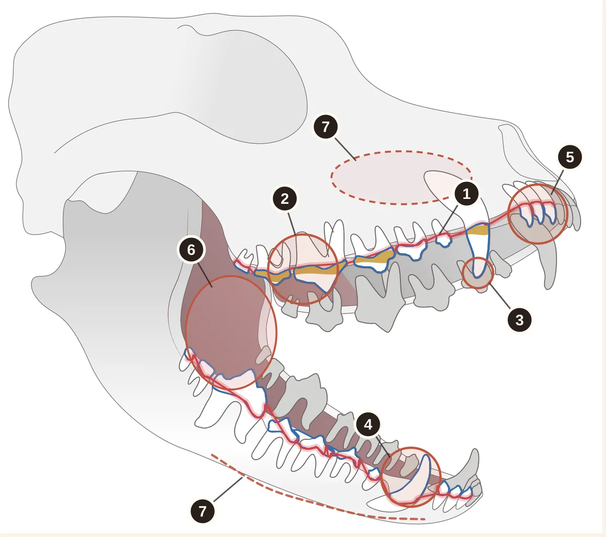
- The gum line. Pink with an even edge. A red line, swelling or blood is a reason for a check-up.
- Plaque and tartar at the gum line. Yellow-brown deposits collect most on the big upper cheek teeth.
- Canine tips and the big upper cheek tooth. Chips, cracks, a pink or dark dot where a tooth has broken.
- Tooth colour. A pink, grey or brown tooth may have a dead pulp.
- Incisors. Are they all there, and are any loose?
- Cheeks, palate and tongue. Ulcers, growths, new dark spots, stuck sticks or bones.
- Face and jaws. Swelling under the eye or along the jaw, asymmetry, pain on touch.
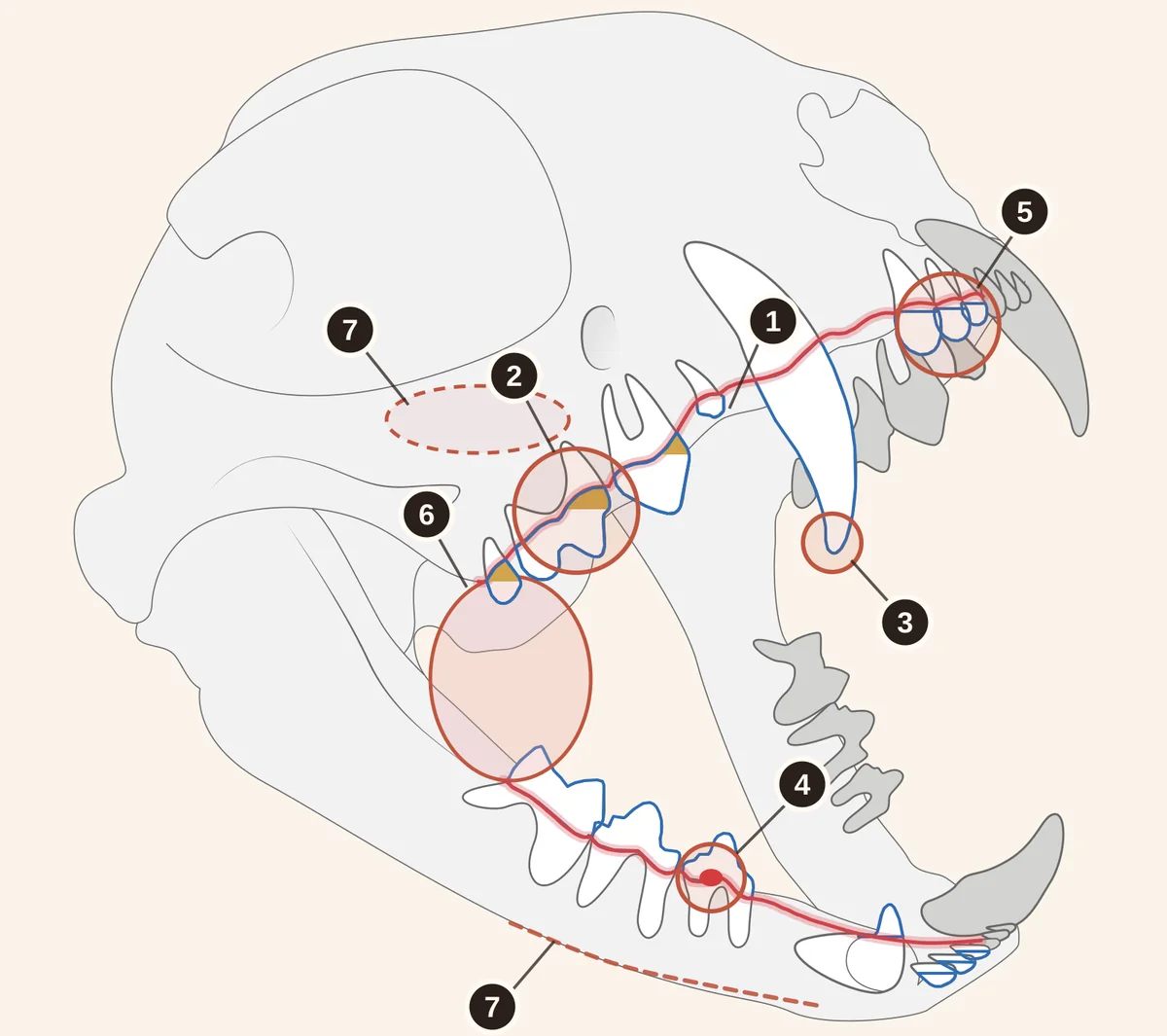
- The gum line. Pink with an even edge. A red line along the teeth, swelling or blood is a reason for a check-up.
- Plaque and tartar at the gum line. Yellow-brown deposits collect most on the big upper cheek teeth.
- Canine tips. Chips, cracks, a pink or dark dot where a tooth has broken.
- A red “bump” at the gum line. The gum seems to creep over the tooth, or a red dot shows at the tooth’s neck — this can be tooth resorption (TR).
- Incisors. Are they all there, and are any loose?
- Back of the mouth, cheeks, tongue. Redness near the last teeth and at the corners of the mouth, ulcers, growths.
- Face and jaws. Swelling under the eye or on the chin, asymmetry, pain on touch.
1. The gum line
Look at the strip of gum along the teeth, especially around the canines and the big side teeth.
- Normal: pink gums (or with natural dark patches), with an even edge that sits snugly against the tooth.
- Worrying: a red line along the teeth, swelling, blood on the gums or on a toy; gums that have receded and exposed the root, or overgrown and are creeping over the tooth. More: Red or bleeding gums.
2. Plaque and tartar at the gum line
Most deposits build up on the big upper side teeth and on the canines, right next to the gums.
- Normal: white or slightly creamy teeth that are smooth and shiny.
- Worrying: soft yellowish plaque that keeps coming back despite brushing, or hard yellow-brown tartar at the gum line. Tartar can no longer be brushed off, and the gums next to it are often inflamed. More: Tartar.
3. Canine tips and the big upper cheek tooth
These are the teeth that break most often: the canines and the big upper cheek tooth (the fourth premolar), the largest tooth in the upper jaw.
- Normal: intact crowns; canine tips that are pointed or evenly worn.
- Worrying: a chip, a sharp broken edge, a pink, red or dark dot in the centre of the break. The dot may mean an exposed pulp (the nerve of the tooth) — then see a vet as soon as possible. In cats the pulp reaches almost to the tip of the canine, so even a small chip often exposes it. More: Broken tooth.
4. Tooth colour
Compare each tooth with its neighbours: what matters is not the shade of all the teeth, but one tooth that stands out.
- Normal: all teeth the same white or cream colour; they may yellow slightly with age.
- Worrying: one tooth that is pink, grey, purple or brown — this happens when the pulp is damaged or has died. More: Discoloured tooth.
- In cats: a pink spot or a “hole” at the gum line, or gum growing over the crown, are typical signs of tooth resorption (TR).
5. Incisors
The incisors are the small front teeth between the canines: both dogs and cats have six at the top and six at the bottom.
- Normal: all incisors present and straight, with firm gums around them.
- Worrying: a gap, a tilted or “longer-looking” tooth with an exposed root, a tooth that moves when your pet licks. In puppies and kittens, a temporary tooth next to a permanent one. Do not wiggle teeth with your finger: the vet checks mobility. More: Loose or missing tooth.
6. Cheeks, palate and tongue
You can see the inside of the cheeks when you draw the lip back. Only look at the palate and tongue when your pet opens its mouth by itself — when yawning or panting after play.
- Normal: pink, smooth lining; dark pigmented patches that have always been there are flat and do not change.
- Worrying: a growth, a sore that does not heal, a new dark spot or one that grows or bleeds; in cats, bright red areas at the back of the mouth. Only an examination can tell what it is, and for a growth only a biopsy. More: Growth or mass on the gums and Dark spot in a dog’s mouth.
- Something stuck: a stick or bone wedged between the teeth or across the palate. Do not pull it out by force — have your pet seen by a vet the same day. More: Bone or stick stuck in the mouth.
7. Face and jaws
Gently run your fingers along the lower jaw and under the eyes on both sides.
- Normal: both sides feel the same, there is no swelling and your pet lets you touch calmly.
- Worrying: swelling under the eye, on the cheek or along the jaw, a sore or draining tract (an opening that leaks pus) under the eye, pain on touch. Swelling under the eye can be linked to a diseased root of an upper tooth. More: Swollen face or swelling under the eye.
THREE MORE SIGNALS
Breath, eating and facial symmetry
You cannot see these on the teeth, but they are often the first hint that something is wrong in the mouth:
- Breath. Smell the breath from a hand’s width away. A mild smell after eating is normal; a persistent rotten smell is not. More: Bad breath.
- Eating. Watch mealtimes for a few days: is your pet chewing on one side, dropping food, eating more slowly or avoiding hard treats; does your cat walk away from the bowl still hungry? More: Changes while eating.
- Symmetry. Step back and look at the face from the front: are both cheeks the same, are both eyes equally open, is saliva dripping from one side, is your pet pawing at its face?
This is how pets show pain even while they keep eating. More in Signs of tooth pain in dogs, Toothache in cats and Myths about dog and cat teeth.
QUICK CHECK
Normal or see the vet: a quick table
Anything in the right-hand column is a reason to book an examination. Anything in the box below is a reason to go as soon as possible.
PHOTOS FOR THE VET
How to photograph the teeth for an online consultation
Photos from the same angles every month are the simplest way to see what has changed. They are also useful for an online consultation if you are not in Kyiv: from photos I can judge how urgent a finding is and suggest what to do next.
Add a few words to the photos: when you noticed the change and whether your pet’s eating has changed. You can book on the consultation page.
- 1Side view — both sidesLift the lip with a finger and photograph the canine and back teeth from the side. Then repeat on the other side.
- 2Front teethGently pull the lips up and down so the incisors, canines and gums are all in the frame.
- 3Back teeth up closeDraw the cheek back towards the corner of the mouth. Tartar and inflammation show best on the large upper teeth.
- 4optionalVideo while eatingYou can also send a short video of your pet while eating.
IMPORTANT
What a home check cannot show
At home you only see the crowns of the teeth and the edge of the gums. The roots, the bone around them, periodontal pockets (gaps between the tooth and the gum) and resorption below the gum line can only be assessed under anaesthesia with dental radiography of all teeth, as part of COHAT. So “all clear” at home does not replace preventive check-ups, and I set how often they are needed individually. You can start with an in-clinic consultation in Kyiv or an online consultation based on your photos.
When to see a vet
- Swelling of the face, under the eye or on the jaw — see a vet as soon as possible.
- A broken tooth with a pink, red or dark dot at the break — as soon as possible.
- Bleeding from the mouth that does not stop, or your pet cannot close its mouth.
- Red or bleeding gums, tartar, a discoloured tooth, a growth, a sore or a new dark spot, persistent bad breath — book an examination without delay.
- Your pet chews on one side, drops food or will not let you touch its face.
What you can do at home
- Take photos from the same angles so you can compare month to month.
- Note your findings on your phone: the date, which tooth and what you saw.
- Get your pet used to having its face touched gradually, with treats: this makes both toothbrushing and vet examinations easier.
- Brush the teeth daily with pet toothpaste — a monthly check does not replace home dental care.
What not to do at home
- Do not force the mouth open or put your fingers between the teeth of a stressed pet.
- Do not press on the gums, wiggle teeth or pick at tartar or a growth.
- Do not try to pull out a temporary tooth or a fragment yourself.
- Do not give painkillers or antibiotics “until the appointment”: ibuprofen, paracetamol (acetaminophen), aspirin, naproxen and other human painkillers are dangerous for dogs and cats, and paracetamol is especially toxic to cats.
Frequently asked questions
A full check with the checklist once a month; during daily brushing you see most of the teeth anyway. While puppies and kittens are changing teeth, look once a week. After treatment, follow the schedule your vet gave you.
Do not insist or restrain your pet by force. Build up gradually: for a few days just touch the face and lips with treats, then lift the lip for a second. If your pet used to allow it and now pulls its head away, this may be a sign of pain — it is better to book an examination.
Plaque is a soft, pale layer at the gum line that comes off with a toothbrush. Tartar is hard, yellow-brown and firmly attached to the tooth, and a toothbrush can no longer remove it. Do not pick at tartar at home: it is removed under anaesthesia together with cleaning below the gum line.
Many dogs have natural dark pigmented patches on the gums, palate or tongue: they are flat, have been there since the dog was young and do not change. A new spot is worth checking, as is one that grows, becomes raised, bleeds or ulcerates. Only an examination can tell what it is, and for a growth only a biopsy.
If you can see a pink, red or dark dot at the break, the pulp (the nerve of the tooth) may be exposed, and your dog should be seen as soon as possible: the sooner treatment starts, the better the chance of keeping the pulp alive. Even a small chip without a dot is best shown to a vet within a few days. Eating normally does not mean the tooth does not hurt.
Sources and professional guidelines
- WSAVA Global Dental GuidelinesGlobal guidelines on veterinary dentistry
- 2019 AAHA Dental Care Guidelines for Dogs and CatsClinical guidelines on dental care
- AVDC Position Statement on Full-Mouth Dental RadiographsPosition on full-mouth dental radiography
- Cornell University — Tooth resorptionOverview of tooth resorption in cats
- American College of Veterinary Surgeons — Oral TumorsOverview of oral tumours in small animals
© dr.vetstomat. Original content.
Copying and publishing text and visual materials without the author's written consent is not permitted, except as allowed by law.