
Four systems on one diagram
The diagrams switch between a dog and a cat: the principle is the same, but a cat has 30 teeth instead of 42, a shorter face and its own weak spots. For the number and shape of the teeth, see How many teeth do dogs and cats have.
Diagram
How the mouth works: four systems
Pick a tab to highlight the nerves, blood vessels, balance of forces or protective barrier on the diagram.
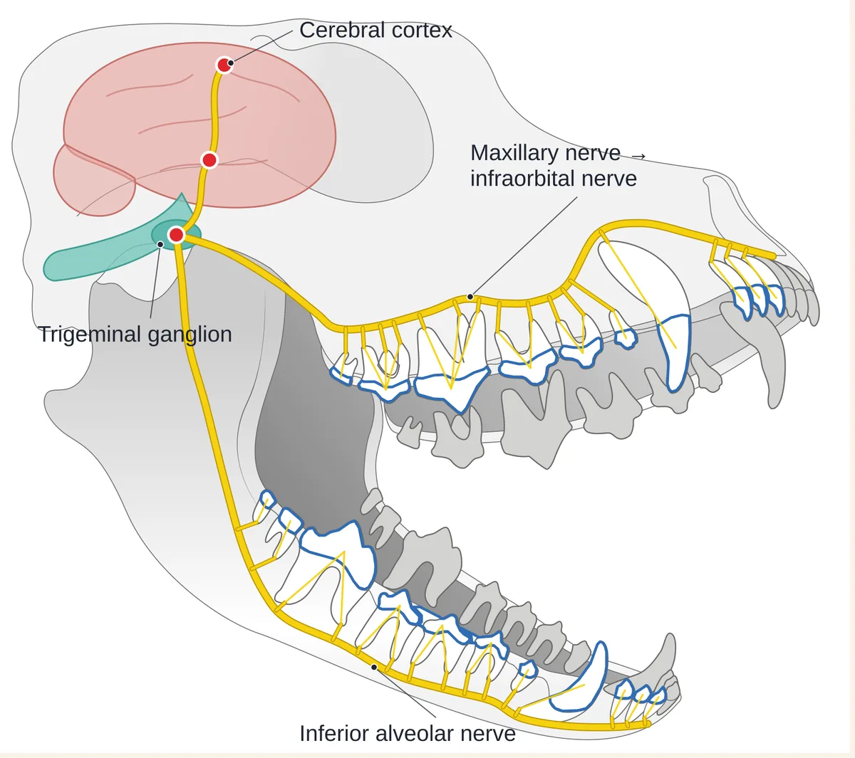
Every tooth has a nerve
Inside every tooth is the pulp, which contains nerve fibres. Sensation from the upper teeth travels along the maxillary and infraorbital nerves, and from the lower teeth along the inferior alveolar nerve in the canal of the lower jaw. They all meet at the trigeminal ganglion and lead to the brain.
What it means for you
A broken or darkened tooth hurts even if your pet eats as usual. Dogs and cats hide pain, so don’t wait for your pet to “show” it.
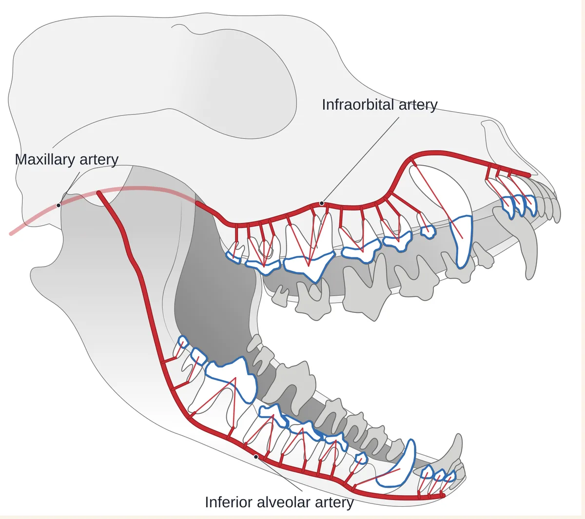
Teeth and gums are fed by the bloodstream
The maxillary artery continues as the infraorbital artery above the roots of the upper teeth. The inferior alveolar artery runs with the nerve inside the lower jaw. A small branch goes to every root, and the gums carry a dense network of tiny vessels.
What it means for you
With periodontitis, bacteria from the pockets around the teeth regularly enter the blood. Periodontal disease is associated with changes in the kidneys, liver and heart, so inflamed gums are not just a mouth problem.
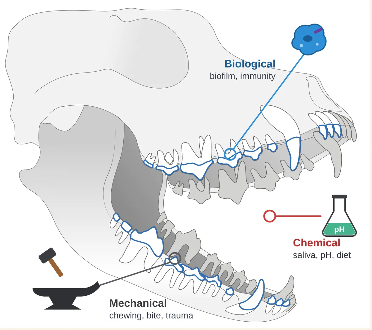
Oral health is a balance of three forces
Biological forces are plaque bacteria against the immune defence of the gums. Mechanical forces are chewing, bite and trauma. Chemical forces are saliva, which rinses and neutralises, together with diet and acidity in the mouth.
What it means for you
If plaque is not removed every day, the balance shifts: the gums become inflamed and plaque hardens into tartar. Daily toothbrushing tips it back towards health.
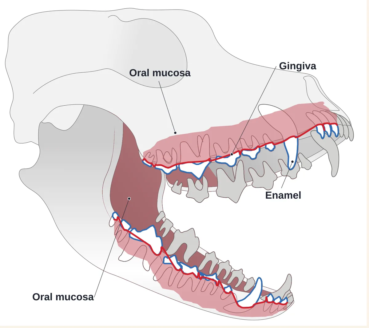
Enamel, gums and mucosa keep bacteria out
Enamel is the hardest tissue in the body, but in dogs and cats it is thin. The gums fit tightly around the neck of the tooth and close the way to the root. The oral mucosa lines the whole mouth and heals quickly.
What it means for you
A chipped tooth, a deep pocket next to a tooth or an ulcer opens a path for bacteria into the tissues and blood. Any such damage is a reason to see a veterinary dentist.
The diagram is simplified and shows a dog’s skull from the side. Switch to the cat to see the feline version.
Nerves: why a tooth hurts even when your pet is eating
Inside every tooth is the pulp: nerves, blood vessels and the cells that nourish the tooth. Sensation from the upper teeth travels along branches of the maxillary nerve, the main one being the infraorbital nerve in its canal above the roots. From the lower teeth it travels along the inferior alveolar nerve in the canal of the lower jaw. Both are branches of the trigeminal nerve, so pain from any tooth reaches the brain.
The pulp is protected by enamel and dentine. When a tooth breaks and the pulp is exposed, or when the pulp becomes inflamed, the tooth hurts. But dogs and cats hardly show dental pain: they keep eating, only chewing on the other side, choosing soft food or playing less with toys. I explain why in Why pets keep eating with painful teeth.
These are the same nerves we block with local anaesthesia during treatment. The pet is under general anaesthesia, and the local block additionally switches off pain in the treated area and helps a calmer recovery.
Blood supply: how infection from the mouth reaches the body
The upper teeth receive blood from the maxillary artery, which continues as the infraorbital artery running above the roots with the nerve of the same name. The lower teeth are supplied by the inferior alveolar artery in the canal of the lower jaw. A small branch goes to every root and enters the pulp through the root tip. The gums and the bone around the teeth carry a dense network of tiny vessels.
This is why the mouth and the rest of the body are closely linked. With periodontitis the gums are inflamed and bleed, and bacteria from periodontal pockets regularly enter the bloodstream. Studies associate periodontal disease in dogs with changes in the kidneys, liver and heart. More in What periodontitis in dogs is and Periodontitis in cats.
Infection can also come from the tooth itself: when the pulp dies, bacteria leave through the root tip into the bone. This causes an abscess, swelling on the cheek or under the eye and a draining tract on the skin. See Can a bad tooth cause swelling under the eye.
Balance: bacteria, bite and saliva
There are always bacteria in the mouth, and that is normal. Health rests on a balance of three forces:
- Biological. Plaque bacteria (biofilm) on one side and the immune defence of the gums on the other. While plaque does not build up, the gums cope with it.
- Mechanical. Chewing, a correct bite and no trauma. A wrong bite injures the gums and palate, and objects that are too hard break teeth.
- Chemical. Saliva rinses away food and neutralises acids, while diet and acidity change the environment. A dog’s mouth is slightly alkaline: this does less to encourage cavities, but tartar forms easily.
Plaque forms on the teeth within hours of brushing, and within a few days it hardens into tartar that a toothbrush can no longer remove. If plaque is not removed, the balance shifts: the gums become inflamed (gingivitis), and over time the inflammation goes deeper, down to the bone — that is periodontitis.
This is why daily brushing with a pet toothpaste is the most effective way to keep the balance. I explain how to get a dog or cat used to it in Home dental care.
The protective barrier: enamel, gums and mucosa
Three layers of protection stand between the bacteria in the mouth and the body’s tissues:
- Enamel is the hardest tissue in the body. But in dogs and cats it is much thinner than in people, and it does not regenerate after the tooth erupts: a chip will not grow back.
- The gums fit tightly around the neck of the tooth and close the way to the root and bone. Healthy gums are pink (or with natural dark patches), with an even margin, not swollen and not bleeding.
- The oral mucosa lines the cheeks, palate, tongue and floor of the mouth. It heals quickly, but ulcers and sores on it are painful.
When the barrier is broken, bacteria get a way in. A chip with exposed dentine or pulp leads to the root, a deep periodontal pocket to the bone, gum recession exposes the root and an ulcer opens the tissues of the cheek or lip. So any such damage is worth showing to a veterinary dentist, even if your pet does not seem bothered.
What is different in cats
Cats have the same system, but there is a disease that breaks the barrier from the inside: tooth resorption (TR). The body itself resorbs the tissues of the tooth, often starting at the gum line or in the root. The defect is painful, but the cat keeps eating, and it is often invisible on the crown until it becomes large.
This is why an examination under anaesthesia with dental radiography matters so much for cats: only the X-ray shows which teeth are already affected. More in Tooth resorption in cats.
- TR 1–2A defect at the gum lineA small pit where tooth meets gum, with red gum tissue growing into it. It may already hurt.
- TR 3The defect reaches the pulpThe tooth hurts, especially when touched or while eating, even if it looks almost intact.
- TR 4The crown breaks downThe crown weakens and may break off. What happens to the root depends on the resorption type, which only an X-ray shows.
- TR 5The crown is goneGum covers the root and the tooth seems to have “fallen out”. Root remnants show only on a radiograph.
How to check that everything is fine in the mouth
Start with an in-clinic consultation: I assess the teeth, gums, bite and mucosa as far as is possible in a conscious pet. If damaged teeth or periodontitis are suspected, the full picture comes from a COHAT procedure under anaesthesia with dental radiography. If you are not in Kyiv, you can start with an online consultation.
When to see a vet
- Swelling of the cheek, lower jaw or under the eye — see a vet as soon as possible.
- A broken tooth, especially with a pink or dark dot at the fracture.
- A tooth has changed colour: pink, grey or brown.
- Red, swollen or bleeding gums, or bad breath that does not go away.
- An ulcer or sore in the mouth that does not heal.
- Your pet chews on one side, drops food from the mouth or avoids having the face touched.
What you can do at home
- Brush the teeth daily with a pet toothpaste: this removes plaque before it turns into tartar.
- Once a month, check the mouth with the home check-up checklist, and glance at the gums during daily brushing.
- Choose chews you can dent with a fingernail and dental products with the VOHC seal.
- Plan preventive check-ups: your vet will suggest how often based on age, breed and the state of the mouth.
What not to do at home
- Do not give hard bones, antlers, stones or hard toys: they break even healthy teeth.
- Do not use human toothpaste: it is not meant to be swallowed.
- Do not scrape tartar off at home with metal tools: it is easy to injure the gums and damage the enamel.
- Do not give antibiotics or painkillers without a vet: they can mask the problem, and ibuprofen and paracetamol are dangerous for pets.
Frequently asked questions
Yes. Dogs’ and cats’ teeth have the same pulp with nerves as ours, and inflammation or an exposed pulp hurts. The difference is that pets hardly show this pain: they keep eating and only change their habits. So a broken or darkened tooth is worth showing to a vet even if your pet does not seem bothered.
With periodontitis, bacteria from inflamed gums regularly enter the bloodstream. Studies associate periodontal disease in dogs with changes in the kidneys, liver and heart. So treating the gums is care not only for the mouth but for the whole body.
Plaque forms within hours of brushing, and within a few days it hardens into tartar that a toothbrush cannot remove. Daily brushing removes plaque before it hardens.
No. Enamel does not regenerate after the tooth erupts, so a chip will not grow back. A small chip without an exposed pulp can often be sealed with a filling; if the pulp is exposed, the tooth needs treatment or extraction. This is decided after an examination and an X-ray.
The principle is the same: nerves, blood vessels, gums and enamel work alike. A cat has fewer teeth — 30 compared with 42 in a dog — and cats often develop tooth resorption, which cannot be seen without an X-ray.
Sources and professional guidelines
- WSAVA Global Dental GuidelinesGlobal guidelines on veterinary dentistry
- 2019 AAHA Dental Care Guidelines for Dogs and CatsClinical guidelines on dental care
- AVDC NomenclatureAVDC nomenclature and terminology
- AVDC: Tooth ResorptionClassification of tooth resorption (TR) by stage and type
© dr.vetstomat. Original content.
Copying and publishing text and visual materials without the author's written consent is not permitted, except as allowed by law.