
Endodontics and root canal treatment
Root canal treatment and vital pulp therapy to preserve traumatised or diseased teeth where clinically indicated.
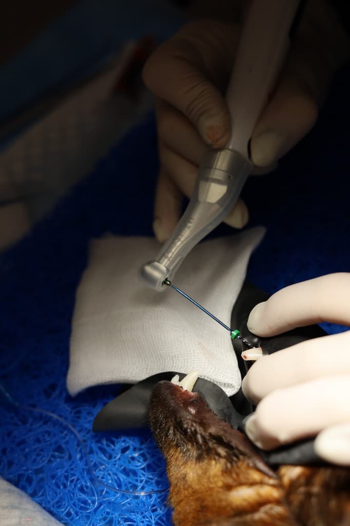
Vital pulpotomy: a chance to keep a freshly fractured tooth alive
Vital pulpotomy (a form of vital pulp therapy) treats a fractured tooth with exposed pulp. I remove only the top layer of pulp at the fracture site — the part that is contaminated and inflamed — and keep the rest of the pulp in the crown and root alive. A medicated material that supports pulp healing is placed over it, and the tooth is sealed with a restoration.
The aim is for the tooth to stay alive and keep working. This matters most in a young dog: living pulp keeps laying down new dentine, the root walls thicken and the tooth grows stronger over the years. If the pulp dies in a young tooth, the root stays thin-walled and more fragile.
Pulpotomy is carried out under inhalational anaesthesia with local dental nerve blocks, with the tooth isolated and strict aseptic technique.
When vital pulpotomy is possible
This approach does not suit every fractured tooth. Timing is decisive: exposed pulp is in immediate contact with oral bacteria, and infection reaches deeper with every day. The sooner treatment follows the injury, the better the chance of keeping the pulp alive — ideally within the first 48 hours.
I make the final decision under anaesthesia, after examining the tooth and taking dental radiographs.
- ✓The fracture is recent and the exposed pulp is still alive
- ✓Most often a young dog with a wide pulp cavity and a root that has not finished developing
- ✓Radiographs show no inflammation in the bone around the root tip and no root fracture
- ✓The tooth can be reliably sealed with a restoration afterwards
How pulpotomy differs from root canal treatment
Both preserve the tooth, but they are used for different pulp conditions. The choice depends on how long ago the injury happened, the animal’s age and the dental radiographs.
Vital pulpotomy
- Recent fracture, living pulp
- Only the top part of the pulp is removed; the rest stays alive
- A young tooth keeps maturing and strengthening
- Follow-up radiographs over time are essential
Root canal treatment
- The pulp is dead or infected, or the fracture is old
- The pulp is removed entirely; the canal is cleaned, disinfected and sealed
- The tooth becomes non-vital but stays functional
- Requires a sufficiently developed root
- Follow-up radiographs are needed here too
The two methods are not rivals: if the pulp dies after pulpotomy, root canal treatment may be the next step. Protecting the crown with a restoration or, when clinically indicated, a crown is planned separately.
Why follow-up radiographs after pulpotomy matter
Pulpotomy is not a one-off procedure but treatment with a monitoring plan. If the pulp dies later, this may not be visible from the outside: the tooth does not necessarily darken, and animals often hide pain. That is why I judge the result on follow-up dental radiographs under anaesthesia.
I usually plan the first check a few months after treatment, and further checks when clinically indicated. We agree the exact timing individually.
- ✓Whether the root keeps developing: thicker walls and a forming root tip
- ✓Whether a protective dentine bridge has formed beneath the medicated material
- ✓Whether there are signs of pulp death or inflammation at the root tip
- ✓Whether the restoration sealing the tooth is intact
Possible outcomes of pulpotomy
The best outcome is that the pulp stays alive, the tooth functions normally and, in a young animal, the root continues to strengthen. Follow-up radiographs confirm this.
The pulp can still die some time after treatment, even when it was done promptly. Root canal treatment is then needed if the root and bone allow, or the tooth is extracted. A tooth like this should not be left untreated: infection spreads from the canal into the bone around the root.
I explain these scenarios in advance so that you understand the monitoring plan before the procedure.
Who this procedure is for
- ✓A tooth fracture with pulp exposure
- ✓A non-vital or infected tooth
- ✓Vital pulp therapy when clinically indicated
- ✓When clinical and radiographic findings allow a functional tooth to be preserved
Symptoms that may indicate this procedure
How diagnosis works
- ✓Clinical examination under anaesthesia
- ✓Dental radiography of the root and surrounding bone tissue
- ✓Assessment of pulp viability
How the procedure works
- ✓Vital pulp therapy where indicated
- ✓Root canal treatment
- ✓Follow-up X-rays
- ✓Crown restoration after root canal treatment
Why anaesthesia is needed
Endodontic procedures are carried out under anaesthesia with local dental nerve blocks.
More on this: anaesthesia in veterinary dentistry.
Technology and equipment
Possible results
- ✓The tooth saved wherever possible
- ✓Pain and the source of infection resolved
- ✓Preservation of tooth function for grasping and breaking up food
What the owner receives after the procedure
- ✓Follow-up advice
- ✓X-ray monitoring over time
- ✓Guidance on how much the tooth can be used
What affects the fee
Cost depends on the tooth, the extent of damage and the scope of endodontic treatment required. Agreed after dental radiography.
Related clinical case
A saved broken tooth in a Jack Russell Terrier
Jack Russell Terrier · 1.3 years
View the tooth-preservation case →Frequently asked questions
No. If the root and surrounding tissue won't allow it, the tooth is extracted. Where conditions are favourable, root canal treatment is considered.
Discolouration is often linked to pulp damage. An examination and X-ray are needed to work out whether the tooth can be saved.
They confirm the canals have been fully filled and that the bone around the root is healing properly.
It is treatment for a recently fractured tooth with exposed pulp: only the contaminated top layer of pulp is removed, a medicated material is placed and the tooth is sealed with a restoration. The rest of the pulp stays alive, so the tooth remains vital and a young tooth keeps getting stronger.
The sooner the better. The best chance of keeping the pulp alive is within the first 48 hours after the fracture; after that bacteria penetrate deeper and the likelihood of success falls. If more time has passed, I assess the tooth on radiographs: root canal treatment or extraction may be options.
The tooth is then treated: root canal treatment is carried out if the root is developed enough and the surrounding bone allows, otherwise the tooth is extracted. This is exactly why follow-up dental radiographs are an essential part of treatment, not a formality.
About dental radiography →Related services
COHAT
COHAT is a comprehensive oral assessment under anaesthesia that includes oral cavity sanitation, periodontal examination, full-mouth dental radiography and evaluation of every tooth.
More about COHAT →Dental Radiography
Full-mouth X-rays reveal roots, bone and changes hidden beneath the gumline.
How dental radiography works →Dental crowns
After trauma, substantial tooth destruction or endodontic treatment, a tooth may need extra protection. One option can be a custom-made dental crown.
More about dental crowns →Restorative & Preventive Dentistry
Composite restorations, correction of enamel defects, Air Flow hygiene and prevention — tailored to individual needs.
More about restorative dentistry →Dental & Jaw Trauma
Fractured and discoloured teeth, jaw injuries — diagnosis, and saving the tooth or extracting it where indicated.
More about dental trauma →© dr.vetstomat. Original content.
Copying and publishing text and visual materials without the author's written consent is not permitted, except as allowed by law.