
Why dogs have dark spots in their mouths
The colour of the lining of the mouth, like the colour of the skin and nose, depends on the pigment melanin. Many dogs have plenty of it in the mouth too: the gums, lips, palate or tongue may be entirely black, grey or bluish, or covered in patches of different shapes. This is a natural feature, not a disease.
In Chow Chows and Shar Peis a blue-black tongue is a breed trait. Spotted gums and tongues are seen in many other breeds and in crossbreeds. Natural pigment has usually been there for a long time: owners often notice it by chance, the first time they look carefully into their dog’s mouth.
Things lying on the teeth rather than in the lining can look dark too: brown or black plaque and tartar at the gum line. This is covered in Yellow, brown and black teeth in dogs. And a dark red or purple patch after an injury may be a bruise: it usually fades within a few days — if it does not, show it to a vet.
How to tell natural pigment from a worrying spot
You cannot make a diagnosis at home, but you can notice whether the spot is changing and what it looks like. Here is a guide:
What is oral melanoma in dogs?
Oral melanoma is a malignant tumour of the pigment cells (melanocytes). In dogs it is one of the most common malignant tumours of the mouth, and it is found more often in middle-aged and older dogs. It can arise on the gums, lips, cheeks, palate or tongue — on dark and on pink lining alike.
Melanoma is serious for two reasons. It grows into the surrounding tissues, including the jawbone, so nearby teeth may become loose. And it can spread (metastasise) to the lymph nodes under the jaw and to the lungs.
In the early stages the tumour may not bother the dog at all: it eats and plays as usual. Bad breath, blood in the saliva, drooling, difficulty eating or swelling appear later. So waiting until the spot “starts causing problems” is not a good idea.
Melanoma can be pink — and a growth is not necessarily melanoma
Some melanomas contain no pigment (they are called amelanotic) and look like a pink or reddish growth, similar to overgrown gum. So a pale colour is no reassurance: a pink lump needs the same check as a dark one.
The reverse is also true: not every growth in the mouth is melanoma. A growth on the gums may be gingival hyperplasia (overgrowth of the gum), an epulis, a papilloma or another tumour, and benign growths can be pigmented too. They cannot be reliably told apart by eye. More on the Growth or lump on the gums page.
How to photograph and monitor a spot at home
Photos are the most reliable way to notice change, because memory lets you down here. This is what I suggest:
- take photos in daylight or with your phone’s torch so that the spot is sharp;
- place a ruler or a coin next to it to show the size;
- photograph from the same side and at the same angle, and note the date;
- repeat the photos during your monthly mouth check and compare them with the earlier ones.
Monitoring is only appropriate for a long-standing flat spot that is not changing. Do not watch a new spot, or one with any of the signs listed above, for months — show it to a vet soon.
How to examine your dog’s mouth calmly and what else to look for is explained in How to check your pet’s mouth at home.
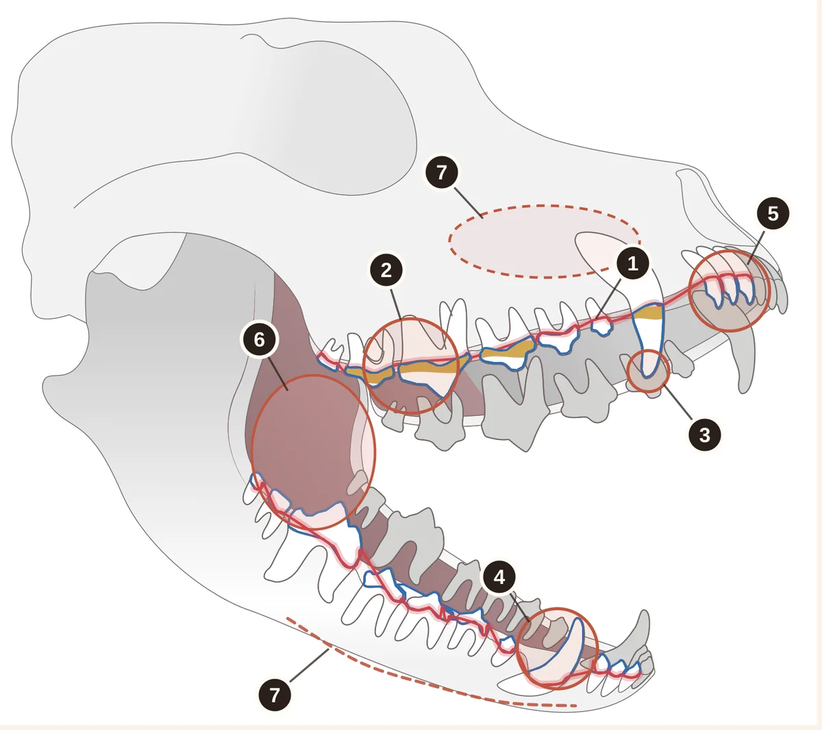
- The gum line. Pink with an even edge. A red line, swelling or blood is a reason for a check-up.
- Plaque and tartar at the gum line. Yellow-brown deposits collect most on the big upper cheek teeth.
- Canine tips and the big upper cheek tooth. Chips, cracks, a pink or dark dot where a tooth has broken.
- Tooth colour. A pink, grey or brown tooth may have a dead pulp.
- Incisors. Are they all there, and are any loose?
- Cheeks, palate and tongue. Ulcers, growths, new dark spots, stuck sticks or bones.
- Face and jaws. Swelling under the eye or along the jaw, asymmetry, pain on touch.
How a suspicious spot is checked
Neither a photo nor the naked eye can tell melanoma from pigment. So the check goes step by step:
- 01
In-clinic consultation
I assess the spot — its size, firmness and edges — the teeth nearby and the lymph nodes under the lower jaw, compare it with your photos and explain what is needed next.
- 02
Examination under anaesthesia and X-rays
Dental radiography shows whether the bone and tooth roots are involved. A CT scan may be needed to plan surgery.
- 03
Biopsy
I take a tissue sample for histopathology, that is, examination of the tissue under a microscope — this is what establishes the diagnosis. A smear of cells (cytology) alone may not be enough.
- 04
Staging
If the tumour is malignant, checks are made to see whether it has spread: the lymph nodes are examined and X-rays or a CT scan of the chest are taken.
- 05
Treatment plan
I explain the results and the treatment options; where needed, the plan is put together with a veterinary oncologist and surgeon.
How oral melanoma in dogs is treated
Treating melanoma is a team effort: a veterinary dentist or surgeon, a veterinary oncologist and, where needed, a radiotherapy specialist. The plan depends on the size and location of the tumour, whether it has spread, and the histopathology results.
Surgery is usually the basis: the tumour is removed together with a margin of healthy tissue, and this not uncommonly means removing part of the jaw. Depending on the stage, the oncologist may suggest radiotherapy, immunotherapy or other methods. How biopsy and removal of growths in the mouth work is described on the Oral tumours page.
It is impossible to promise an outcome in advance, and I do not do so. But a small tumour is generally easier to remove with an adequate margin than a large one, so it is best not to delay with a suspicious spot.
When to see a vet
- The spot or growth bleeds, grows quickly, or the jaw or face has swollen — an examination within days.
- A new dark spot in an adult or older dog — without waiting for it to get bigger.
- The spot has become raised, changed shape or colour, or has uneven edges.
- Teeth next to the spot are loose or have shifted, or there is bad breath or blood in the saliva.
- Any new lump in the mouth — dark or pink, even if small and painless.
- Enlarged lumps under the lower jaw (lymph nodes).
What you can do at home
- Photograph the spot in daylight next to a ruler or coin and note the date.
- Repeat the photos under the same conditions during your monthly mouth check so you can compare.
- Show a long-standing spot at a routine check-up so the vet can assess it and record it in the notes.
- Unsure how urgent it is? Send photos for an online consultation — but a photo does not replace an examination or a biopsy.
What not to do at home
- Do not scrape, prick, cauterise or try to cut off the spot or growth.
- Do not apply ointments, gels or “home remedies”: they do not treat tumours and only delay the diagnosis.
- Do not wait for the spot to “grow noticeably” to be sure: a small growth is easier to remove with a margin of healthy tissue.
- Do not compare it with photos from the internet: growths that look alike can be completely different in nature.
- Do not give human painkillers: ibuprofen, paracetamol and others are dangerous for dogs.
Frequently asked questions
Yes, in many dogs the gums, lips, palate or tongue are partly or completely dark with natural pigment. This is normal if the pigment is flat, smooth, painless and unchanging over time. New, raised or changing spots, ulcers or bleeding are a concern.
Usually not: spotted tongues are seen in dogs of many breeds and in crossbreeds, and Chow Chows and Shar Peis naturally have blue-black tongues. If a spot on the tongue is new, raised, bleeding or ulcerated, show it to a vet.
No. From a photo I can judge how urgently an examination is needed, but not what the spot is. Only histopathology of a tissue sample — a biopsy — reliably distinguishes melanoma from a benign growth or natural pigment.
Show it at your next routine check-up so the vet can assess it and record it in the notes, and photograph it at home next to a ruler for comparison. If the spot starts to change — growing, becoming raised or bleeding — do not wait for the routine check-up.
Yes. Some melanomas contain no pigment and look like a pink or reddish growth, similar to overgrown gum. So a pale colour does not mean a growth is harmless: it needs checking just like a dark one.
There are treatments, and the plan is made after a biopsy and staging. Usually this means surgery to remove the tumour with a margin of healthy tissue, sometimes with part of the jaw, plus radiotherapy or other methods suggested by a veterinary oncologist. The outlook depends on the size, location and spread of the tumour, so no outcome can be promised in advance.
Ginger and tortoiseshell or calico cats not uncommonly develop flat black freckles on the lips, gums, eyelids and nose as they age — this is benign pigmentation (lentigo). But a raised spot, a growth, an ulcer or bleeding in a cat’s mouth should also be shown to a vet.
Sources and professional guidelines
- American College of Veterinary Surgeons — Oral TumorsOverview of oral tumours in small animals
- Cornell University — Oral cavity tumorsOverview of oral cavity tumours
- Cornell University — Diagnosis and staging: principles of tumor biopsiesPrinciples of tumour biopsy
- Simões et al. — Cytological and histopathological diagnoses of oral lesions in dogs and cats, 2025Comparison of cytology and histology of oral lesions
- WSAVA Global Dental GuidelinesGlobal guidelines on veterinary dentistry
© dr.vetstomat. Original content.
Copying and publishing text and visual materials without the author's written consent is not permitted, except as allowed by law.