
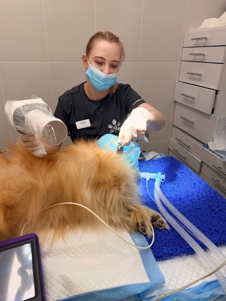

What a dental X-ray shows
X-rays allow the roots to be assessed, along with the bone around them, and reveal abscesses, root fractures and resorption.
In cats, X-rays are especially important for spotting resorption, which often starts below the gumline.
- Condition of the roots and pulp chamber
- Level of bone support around the tooth
- Hidden fractures and infections
When an X-ray is essential
During any dental treatment under anaesthesia, an X-ray is the standard of modern practice. It helps decide whether to save a tooth, treat the root canal, or extract it.
Safety of the images
Modern veterinary dental X-ray equipment delivers a minimal radiation dose. Images are taken under anaesthesia, while the pet is still — this ensures image quality.
When to see a vet
- Dental treatment under anaesthesia is planned — an X-ray is part of the standard protocol
- There are symptoms (pain, swelling, a darkened tooth) but a visual examination doesn't provide an answer
- You need to decide whether a tooth can be saved — without an X-ray the answer would only be a guess
What not to do at home
- Don't refuse an X-ray out of fear of radiation without discussing it with your vet
- Don't assume a visual examination is enough for a full assessment
- Don't expect an accurate diagnosis "by eye" — hidden disease is common
Frequently asked questions
Usually a full set of dental images of the whole mouth is taken, so every tooth can be assessed systematically.
You can do a surface clean only, but a thorough diagnosis and evidence-based treatment aren't possible without images.
Sources and professional guidelines
- WSAVA Global Dental Guidelines ↗Глобальні настанови з ветеринарної стоматології
- AAHA Dental Care Guidelines for Dogs and Cats ↗Клінічні настанови з дентального догляду
© dr.vetstomat. Original content.
Copying and publishing text and visual materials without the author's written consent is not permitted, except as allowed by law.