
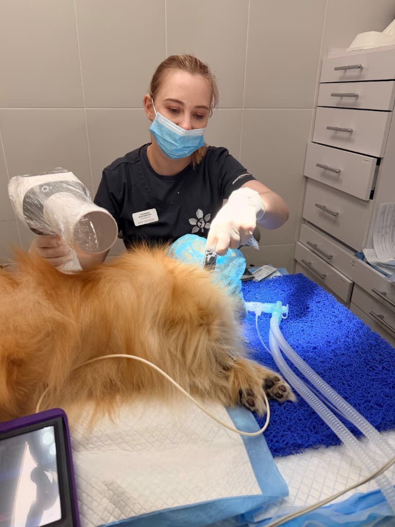
What a dental X-ray shows
A dental X-ray lets me see what is physically out of reach in an examination: the roots of the teeth, the pulp chambers, the level of bony support, root fractures, abscesses, root remnants left after earlier extractions, bone defects and tooth resorption (TR) in cats.
In dogs I regularly find root infections in teeth that look only slightly darkened from outside. In cats, X-rays are indispensable for diagnosing TR — resorption that often begins below the gumline and progresses inside the tooth.
- The condition of the roots and pulp chamber of every tooth
- The level of bony support and the depth of periodontal pockets
- Hidden fractures, cracks and infections
- Tooth resorption (TR) in cats — stage and type
- Root remnants and bone damage after injuries
When an X-ray is essential
In any dental treatment under anaesthesia — as part of the COHAT protocol — full-mouth dental radiography is the standard. I take a set of images to assess all the teeth systematically, not only those that look “suspicious” from outside.
An X-ray is also needed separately when there are symptoms — pain, swelling, a darkened tooth, bleeding — but the external examination gives no answer. It is also needed before deciding whether to keep a broken tooth, treat the canals or extract: without an image the conclusion will be approximate.
Typical “hidden” findings on X-rays
The most common surprises on images: loss of bony support around a tooth that looks “normal” from outside; a root fracture with no visible chip of the crown; infection at the root tip with an abscess; TR at a stage when the tooth still looks intact; a root remnant after an incomplete extraction.
Without X-rays these problems stay undetected — the animal goes on living with chronic pain, and the owner cannot understand why the symptoms do not disappear after “cleaning”.
Why a pet may keep eating even when the X-ray shows disease
I regularly see marked damage on images in animals that keep eating. Dogs and cats adapt: they chew on one side, choose soft food, swallow quickly. This does not mean there is no pain — it means that an external examination and behaviour do not reflect the real condition.
That is why I insist on a full set of images, not a “spot” X-ray of a single tooth. The disease is often multiple.
Safety of dental images
Modern dental X-ray equipment for animals gives a minimal radiation dose — much lower than many medical investigations. The images are taken under anaesthesia while the animal is still, which ensures image quality and reduces the need for repeat images.
If an owner is worried about radiation, I explain that the risk from an untreated root infection or progressing periodontitis considerably outweighs the risk from dental images.
What the owner should do
If you have been offered a dental procedure, ask whether dental X-rays are part of the protocol. If you are told X-rays are not needed “to save money”, that is a reason to discuss how complete the diagnosis will be.
At the consultation I explain which images I plan and how they will affect the treatment plan. After the procedure you receive the images in your report — a document you can come back to at future check-ups.
When to see a vet
- Dental treatment under anaesthesia is planned — X-rays are part of the standard protocol
- There are symptoms (pain, swelling, a darkened tooth) but the external examination gives no answer
- You need to decide whether a tooth can be kept — without X-rays the conclusion will be approximate
- Symptoms persisted after “cleaning” — hidden root disease is possible
- The pet chews on one side but the teeth “look normal”
- You need an assessment after an injury to a tooth
- Resorption (TR) is suspected in a cat — X-rays are essential
- Extraction or canal treatment is planned — without an image the root cannot be assessed
What not to do at home
- Do not decline X-rays out of fear of radiation without discussing it with the vet
- Do not assume an external examination is enough for a full assessment
- Do not expect an accurate diagnosis “by eye” — hidden disease is common
- Do not accept “cleaning without X-rays” as complete treatment
- Do not interpret the images yourself — a professional assessment is needed
Frequently asked questions
Yes, usually a full set of dental images of the mouth is taken — several images per quadrant, to cover all the teeth systematically. I do not limit myself to the “suspicious” tooth.
Only surface hygiene can be done, but a complete diagnosis and well-founded treatment are impossible without images. I do not recommend that approach.
No, the images are taken under anaesthesia. The animal feels no discomfort. The sensor is placed in the mouth for a few seconds.
Yes, X-rays are what allow the stage and type of TR to be determined, the condition of the root to be assessed and treatment to be planned. An external examination is not enough for TR.
The dose is minimal. For older animals the risk of untreated dental disease is considerably higher than the risk from the images. The vet assesses this individually.
Sources and professional guidelines
- WSAVA Global Dental GuidelinesGlobal guidelines for veterinary dentistry
- AAHA Dental Care Guidelines for Dogs and CatsClinical guidelines for dental care
- AVDC NomenclatureAVDC nomenclature and terminology
© dr.vetstomat. Original content.
Copying and publishing text and visual materials without the author's written consent is not permitted, except as allowed by law.